A single gene can encode not one but dozens of distinct proteins through alternative splicing of its pre-mRNA, and the choice of which splice variant a cell produces is itself a tightly regulated decision. External signals — hormones, growth factors, cytokines, and metabolites — must traverse the space between the cell surface and the nucleus before they can alter transcription, and the signalling cascades that accomplish this transmission are themselves subject to elaborate control. After transcription, the fate of an mRNA molecule is far from settled: its stability in the cytoplasm, its localization within the cell, and the efficiency with which ribosomes translate it all constitute independent regulatory layers, each capable of producing dramatic changes in protein output without altering transcription at all.
Perhaps the most conceptually important development in modern gene regulation is the recognition that individual regulatory interactions do not operate in isolation. Transcription factors regulate each other, forming networks whose topology determines the dynamic behaviour of the cell. Feed-forward loops filter transient signals from sustained ones. Bistable switches lock cells into one of two alternative fates. Oscillatory circuits generate rhythmic gene expression that drives processes from somite formation to the circadian clock. These network motifs, recurring across organisms from bacteria to humans, reveal a deep logic underlying the apparent complexity of gene regulation.
The material traces the regulatory journey from a freshly transcribed pre-mRNA through the splicing machinery, follows external signals as they propagate from receptor to nucleus, examines the post-transcriptional controls that shape the proteome without touching the genome, and culminates in the network-level logic that converts thousands of individual regulatory interactions into coherent cellular decisions. Along the way, it will become clear that the genome is not merely read but interpreted, and that interpretation depends on layers of control far richer than the linear sequence of DNA could suggest.
1. Alternative Splicing and the Expansion of the Proteome
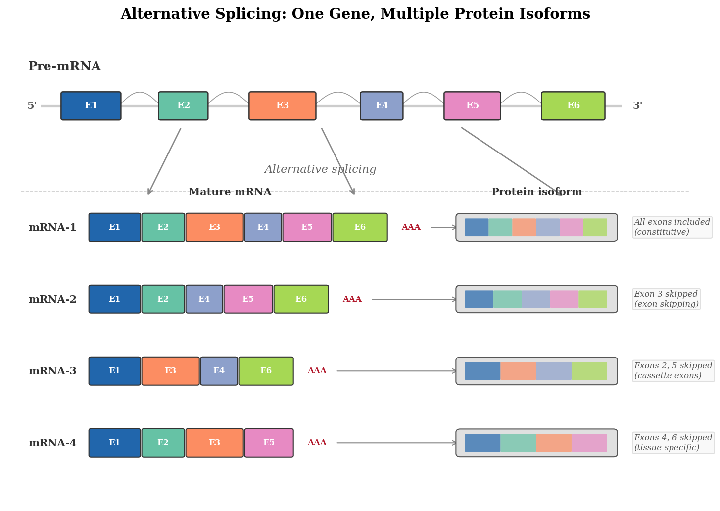
The human genome contains approximately 20,000 protein-coding genes, yet the human proteome — the full set of proteins a human body can produce — numbers well over 100,000 distinct species. The discrepancy between gene number and protein number was one of the most striking revelations of the post-genomic era, and its resolution lies in a molecular process called alternative splicing. When RNA polymerase II transcribes a gene, the resulting pre-mRNA contains both exons (the sequences that will appear in the mature mRNA) and introns (intervening sequences that must be removed). In constitutive splicing, every exon is included and every intron is excised, producing a single mature mRNA from a given gene. In alternative splicing, the splicing machinery selects different combinations of exons, producing multiple distinct mRNAs from the same primary transcript.
The scale of alternative splicing in the human genome is remarkable. Large-scale RNA sequencing studies have shown that over 95 percent of human multi-exon genes undergo alternative splicing, meaning that the vast majority of genes can produce more than one protein isoform. Some genes are extraordinarily versatile: the Drosophila gene Dscam, involved in neuronal wiring, can theoretically produce over 38,000 distinct mRNA isoforms through combinatorial use of its alternatively spliced exon clusters, more than twice the number of genes in the entire Drosophila genome. While human genes rarely approach this extreme, many important genes produce dozens of functionally distinct isoforms whose relative abundances vary dramatically across tissues, developmental stages, and physiological states.
The molecular machinery that executes splicing is the spliceosome, a massive ribonucleoprotein complex composed of five small nuclear RNAs (U1, U2, U4, U5, and U6) and over 150 associated proteins. The spliceosome assembles de novo on each intron, recognising splice sites through a series of RNA-RNA base-pairing interactions. The 5' splice site (at the upstream end of the intron), the branch point (an adenosine residue within the intron), and the 3' splice site (at the downstream end) must all be correctly identified for accurate excision. The chemistry of splicing involves two sequential transesterification reactions: the 2'-hydroxyl of the branch-point adenosine attacks the phosphodiester bond at the 5' splice site, forming a lariat structure, and then the free 3'-hydroxyl of the upstream exon attacks the phosphodiester bond at the 3' splice site, joining the two exons and releasing the intron lariat.
Patterns of Alternative Splicing and Their Functional Consequences
Alternative splicing takes several mechanistically distinct forms, each with different consequences for the resulting protein. The most common is exon skipping (also called cassette-exon inclusion), in which an entire exon is either included or excluded from the mature mRNA. In the Fas receptor gene, inclusion of exon 6 produces the membrane-bound pro-apoptotic isoform, while skipping of exon 6 produces a secreted decoy receptor that inhibits apoptosis — a single exon-skipping event that determines whether a cell lives or dies. Alternative 5' splice site and alternative 3' splice site selection extend or shorten individual exons by choosing between two competing splice sites within the same intron-exon boundary, often adding or removing a few amino acids from a protein domain and thereby altering binding affinity or enzymatic activity. Intron retention, in which an intron is left unspliced in the mature mRNA, is particularly prevalent in plants and is increasingly recognised as a regulatory mechanism in mammalian cells, where retained introns often introduce premature stop codons that target the transcript for nonsense-mediated decay. Mutually exclusive exon selection, where exactly one of two or more adjacent exons is included, allows genes to produce structurally related but functionally distinct protein variants — a strategy exploited by ion channels and cell-adhesion molecules to generate the precise biophysical diversity required for neural circuit assembly. The combinatorial use of these splicing patterns across multiple positions within a single gene is what allows Dscam to generate its remarkable 38,000 isoforms and what makes the splicing code a powerful amplifier of genomic information.
The Regulatory Logic of Splice-Site Selection
If the spliceosome simply recognised every splice site with equal efficiency, alternative splicing would not occur. The key to regulated splicing is that splice sites vary in their strength — the degree to which their sequences match the consensus recognised by spliceosomal components — and that trans-acting factors modulate splice-site recognition. Two major families of splicing regulators govern these decisions: SR proteins (serine/arginine-rich proteins) and heterogeneous nuclear ribonucleoproteins (hnRNPs). SR proteins generally promote exon inclusion by binding to exonic splicing enhancer sequences and recruiting spliceosomal components to nearby splice sites. hnRNPs generally promote exon skipping by binding to exonic or intronic splicing silencer sequences and sterically blocking spliceosome assembly.
The relative concentrations and activities of SR proteins and hnRNPs vary between cell types and developmental stages, creating tissue-specific splicing programmes. The neural-specific SR protein nPTB (neural polypyrimidine tract-binding protein), for example, is expressed predominantly in the brain and promotes the inclusion of neuron-specific exons in dozens of pre-mRNAs, contributing to the distinctive protein repertoire of neurons. Conversely, the ubiquitous hnRNP PTB represses these same exons in non-neural tissues. The switch from PTB to nPTB during neural differentiation is therefore a master regulatory event that reprogrammes the splicing landscape of the differentiating cell.
Alternative splicing is not merely a way to increase protein diversity; it is a regulatory mechanism in its own right. Some alternative exons introduce premature stop codons into the mRNA, targeting the transcript for degradation by a quality-control pathway called nonsense-mediated mRNA decay. This coupling of splicing to mRNA degradation, known as regulated unproductive splicing and translation (RUST), allows cells to control gene expression quantitatively by adjusting the fraction of transcripts that include or exclude the poison exon. The abundance of a functional protein can thus be tuned without changing the rate of transcription or the activity of any transcription factor — a post-transcriptional rheostat embedded within the gene itself.
Splicing in Disease and as a Therapeutic Target
The clinical importance of alternative splicing became dramatically clear with the molecular dissection of spinal muscular atrophy (SMA), a devastating neurodegenerative disease caused by loss of the SMN1 gene. Humans possess a nearly identical paralog, SMN2, but a single nucleotide difference in exon 7 of SMN2 weakens an exonic splicing enhancer, causing the spliceosome to skip exon 7 in approximately 90 percent of SMN2 transcripts. The resulting truncated protein is unstable and non-functional. Because exon 7 skipping is a splicing decision rather than a genomic deletion, it is in principle reversible. The antisense oligonucleotide drug nusinersen (Spinraza), approved in 2016, binds to an intronic splicing silencer downstream of exon 7 in SMN2 pre-mRNA, blocking the binding of a repressive hnRNP and redirecting the spliceosome to include exon 7. The result is a dramatic increase in full-length, functional SMN protein, and the clinical outcomes in treated infants have been transformative — children who would otherwise never sit unaided are walking.
The success of nusinersen illustrates a broader principle: because splicing is a decision point in gene expression, it is a druggable decision point. Antisense oligonucleotides, small molecules that modulate spliceosome activity, and CRISPR-based approaches to edit splice-site sequences are all being explored as therapeutic strategies for diseases ranging from Duchenne muscular dystrophy (where exon skipping can restore the reading frame of a mutated dystrophin gene) to certain cancers (where aberrant splicing of apoptotic regulators contributes to tumour survival). The recognition that the splicing code is as therapeutically accessible as the genetic code has opened a new chapter in molecular medicine.
2. Signal Transduction: How External Cues Reach the Genome
A cell in a multicellular organism does not regulate its genes in isolation. It is continuously bathed in a complex mixture of signalling molecules — hormones, growth factors, cytokines, neurotransmitters, and metabolites — that carry information about the organism's developmental state, physiological needs, and environmental conditions. For this information to alter gene expression, it must be transmitted from the cell surface to the nucleus, a journey that involves a cascade of molecular interactions collectively called signal transduction. The signalling pathway is the molecular wire that connects an extracellular cue to a transcriptional response, and understanding its architecture is essential to understanding how gene regulation operates in context.
The canonical architecture of a signalling pathway begins with a receptor at the cell surface. Receptor tyrosine kinases (RTKs), for example, are transmembrane proteins whose extracellular domains bind specific ligands — epidermal growth factor (EGF), fibroblast growth factor (FGF), insulin, and many others — and whose intracellular domains possess enzymatic activity. Ligand binding induces receptor dimerisation, which activates the intracellular kinase domains through trans-autophosphorylation. The phosphorylated tyrosine residues on the receptor's cytoplasmic tail then serve as docking sites for adaptor proteins containing SH2 (Src homology 2) or PTB (phosphotyrosine-binding) domains. These adaptor proteins recruit and activate downstream effectors, initiating a chain of protein-protein interactions and enzymatic modifications that propagates the signal toward the nucleus.
One of the best-characterised signalling cascades is the RAS-MAPK pathway. Upon activation of an RTK, the adaptor protein GRB2 recruits the guanine nucleotide exchange factor SOS, which activates the small GTPase RAS by promoting the exchange of GDP for GTP. Active RAS recruits and activates the serine/threonine kinase RAF, which phosphorylates and activates MEK, which in turn phosphorylates and activates ERK (extracellular signal-regulated kinase). ERK then translocates to the nucleus, where it phosphorylates transcription factors such as ELK1, c-FOS, and c-MYC, altering their activity and thereby changing the transcriptional programme of the cell. The entire cascade, from ligand binding at the cell surface to transcription factor activation in the nucleus, can occur within minutes.
Amplification, Specificity, and the Problem of Cross-Talk
The multi-step architecture of signalling pathways is not merely a relay; it provides opportunities for amplification, integration, and specificity that a simple one-step mechanism could not achieve. At each level of the cascade, a single activated kinase can phosphorylate multiple substrate molecules, so the signal is amplified as it propagates. A single activated RAS molecule can activate multiple RAF molecules; each RAF molecule can activate multiple MEK molecules; each MEK molecule can activate multiple ERK molecules. The result is that a small number of ligand-receptor binding events at the cell surface can produce a large transcriptional response in the nucleus.
Specificity presents a more challenging problem. The RAS-MAPK pathway is activated by dozens of different growth factors through dozens of different RTKs, yet the cellular response — proliferation versus differentiation versus survival — differs depending on which growth factor is involved, in which cell type, and at which developmental stage. How does a single pathway produce diverse outputs? Part of the answer lies in signal duration: transient activation of ERK (lasting minutes) tends to promote proliferation, while sustained activation (lasting hours) can trigger differentiation, as Peter Marshall and colleagues demonstrated in the classic PC12 cell system where nerve growth factor (NGF) and EGF produce opposite outcomes despite both activating RAS-MAPK. Part of the answer lies in combinatorial signalling: a cell typically receives multiple signals simultaneously, and the transcriptional response integrates the combined activities of multiple pathways converging on the same promoters and enhancers. And part of the answer lies in scaffold proteins — large, multidomain proteins that physically tether specific combinations of kinases together, ensuring that a particular receptor activates a particular subset of downstream effectors while preventing cross-activation of unrelated pathways.
The JAK-STAT pathway illustrates a different signalling architecture with a more direct route to transcription. Cytokine receptors activate Janus kinases (JAKs), which phosphorylate signal transducers and activators of transcription (STATs). Phosphorylated STATs dimerise, translocate to the nucleus, and bind DNA directly, activating specific target genes. The relative simplicity of this pathway — only one phosphorylation step separates receptor activation from transcription factor binding — means that the JAK-STAT response is exceptionally rapid, consistent with its role in immune signalling where speed is critical. However, the same simplicity limits the opportunities for signal modulation, which is why the JAK-STAT pathway is subject to elaborate negative regulation by SOCS (suppressor of cytokine signalling) proteins, protein tyrosine phosphatases, and PIAS (protein inhibitor of activated STAT) proteins.
Nuclear Hormone Receptors: When the Signal Is the Transcription Factor
Not all signalling molecules require transmembrane receptors. Steroid hormones — cortisol, oestrogen, testosterone, aldosterone, and their relatives — are small, hydrophobic molecules that can diffuse directly across the plasma membrane and enter the cell. Once inside, they bind to intracellular receptors belonging to the nuclear hormone receptor superfamily. In the absence of ligand, these receptors are typically sequestered in the cytoplasm in complex with heat-shock proteins (such as HSP90) that hold them in an inactive conformation. Ligand binding triggers a conformational change that releases the receptor from the chaperone complex, exposes a nuclear localisation signal, and allows the receptor to translocate to the nucleus. There, the ligand-bound receptor dimerises (often as a homodimer) and binds to specific DNA sequences called hormone response elements (HREs) in the regulatory regions of target genes, recruiting coactivator complexes that include histone acetyltransferases and components of the Mediator complex, and directly stimulating transcription.
The nuclear hormone receptor mechanism collapses the distinction between signal and transcription factor. The receptor is both: it senses the presence of the hormone and it binds DNA to activate (or repress) target genes. This dual identity means that nuclear hormone receptor signalling is exceptionally direct — there is no multi-step kinase cascade, no amplification through intermediary enzymes. The trade-off is that the response is slower to initiate (the ligand must diffuse to the nucleus and the receptor-DNA complex must recruit the transcriptional machinery) but potentially longer-lasting, because the receptor remains bound to DNA as long as the ligand is present. This makes nuclear hormone receptor signalling well-suited to physiological processes that require sustained, coordinated changes in gene expression across many target genes simultaneously — as in the metabolic and developmental programmes controlled by thyroid hormone, retinoic acid, and the sex steroids.
The pharmacological importance of nuclear hormone receptors is difficult to overstate. Tamoxifen, the selective oestrogen receptor modulator used in breast cancer therapy, works by binding the oestrogen receptor and inducing a conformation that recruits corepressors rather than coactivators, converting the receptor from a transcriptional activator into a transcriptional repressor at its target genes. Dexamethasone, a synthetic glucocorticoid, activates the glucocorticoid receptor to suppress inflammatory gene expression. These drugs work because they intervene precisely at the point where signal transduction meets transcription — the ligand-binding pocket of a nuclear hormone receptor — and because the specificity of receptor-ligand interaction ensures that only the target pathway is modulated.
Negative Regulation and the Termination of Signalling
A signalling pathway that could not be shut off would be as dangerous as one that could not be turned on. Cells deploy multiple mechanisms to ensure that every signalling event is self-limiting. Receptor tyrosine kinases are internalised by clathrin-mediated endocytosis shortly after ligand binding, and the internalised receptor-ligand complex is either recycled to the surface or targeted to lysosomes for degradation — a process that physically removes the signalling platform from the plasma membrane. Protein phosphatases counterbalance kinase activity at every level of the cascade: SHP1 and SHP2 dephosphorylate receptor tyrosine residues, DUSP (dual-specificity phosphatase) family members inactivate ERK and other MAP kinases, and PP2A dephosphorylates components of the PI3K-AKT pathway. The RAS GTPase itself is a molecular timer, hydrolyzing its bound GTP to GDP (accelerated by GTPase-activating proteins such as NF1) and thereby returning to the inactive state. Negative feedback loops add another layer: ERK phosphorylates and inhibits SOS, the very exchange factor that activated RAS in the first place, creating a circuit-level brake on the entire cascade. In the JAK-STAT pathway, the STAT target genes include SOCS proteins that bind back to the activated receptor and block further JAK phosphorylation. The elaborate architecture of signal termination ensures that transcriptional responses are proportional to the stimulus and decay when the stimulus is removed — a prerequisite for a cell that must interpret a continuous, changing stream of extracellular information.
3. Post-Transcriptional Regulation: mRNA Stability, Localisation, and Translational Control
The decision to transcribe a gene is only the first of many steps that determine how much protein a cell ultimately produces from that gene. Once an mRNA has been transcribed, processed, and exported from the nucleus, it enters a cytoplasmic environment where its fate is controlled by a distinct set of regulatory mechanisms that operate entirely independently of the transcriptional machinery. The abundance of a protein in a cell is determined not only by how fast the gene is transcribed but also by how long the mRNA persists before it is degraded, how efficiently ribosomes translate it, and where within the cell the translation occurs. These post-transcriptional controls constitute a parallel regulatory layer that allows cells to respond rapidly to changing conditions — far faster than the minutes to hours required to alter transcription, export new mRNAs, and accumulate new protein from freshly initiated transcripts.
The stability of an mRNA in the cytoplasm varies enormously between transcripts. Some mRNAs, such as those encoding ribosomal proteins and other housekeeping factors, have half-lives of many hours or even days. Others, particularly those encoding transcription factors, signalling molecules, and inflammatory mediators, have half-lives of minutes. The difference often lies in sequence elements within the 3' untranslated region (3' UTR) of the mRNA. AU-rich elements (AREs), consisting of repeated AUUUA pentamers or related sequences, are among the best-characterised destabilising elements. ARE-binding proteins such as tristetraprolin (TTP) recruit the cellular mRNA decay machinery — deadenylases that shorten the poly(A) tail, the exosome complex that degrades the mRNA body in the 3'-to-5' direction, and the decapping complex that exposes the 5' end to 5'-to-3' exonuclease attack. Conversely, stabilising proteins such as HuR can bind the same or overlapping elements and protect the mRNA from degradation. The balance between destabilising and stabilising factors at the 3' UTR determines the steady-state half-life of the transcript.
The regulation of mRNA stability is not static; it responds dynamically to cellular conditions. The mRNA encoding the inflammatory cytokine tumour necrosis factor alpha (TNF-alpha), for example, contains multiple AREs in its 3' UTR and is normally extremely unstable, with a half-life of approximately 15 minutes. Upon activation of macrophages by bacterial lipopolysaccharide (LPS), signalling through the p38 MAPK pathway phosphorylates and inactivates TTP, stabilising the TNF-alpha mRNA and allowing its accumulation. The result is a rapid, many-fold increase in TNF-alpha protein production that occurs largely through post-transcriptional stabilisation rather than through increased transcription. This mechanism enables the immune system to respond explosively to infection while maintaining tight control under resting conditions.
P-Bodies, Stress Granules, and Cytoplasmic RNA Compartments
The enzymes and regulatory factors that control mRNA decay and translational repression are not uniformly distributed in the cytoplasm. Instead, they concentrate in discrete, membrane-less cytoplasmic granules whose formation is driven by liquid-liquid phase separation of RNA-binding proteins containing intrinsically disordered, low-complexity domains. Processing bodies (P-bodies) are constitutive structures enriched in decapping enzymes (DCP1/DCP2), the 5'-to-3' exonuclease XRN1, deadenylases, and translational repressors. mRNAs that have been targeted for silencing — whether by miRNA-loaded RISC, ARE-binding proteins, or other decay factors — accumulate in P-bodies, where they can be either degraded or stored in a translationally repressed state and later returned to the translating pool if conditions change. Stress granules form transiently when global translation is inhibited, as occurs during the eIF2-alpha phosphorylation response described below. They contain stalled 48S pre-initiation complexes, poly(A)-binding protein, the RNA helicase eIF4A, and a variety of RNA-binding proteins including TIA-1 and G3BP1. Stress granules are thought to serve as triage centres where mRNAs are sorted: housekeeping transcripts are sequestered and protected, while stress-response mRNAs (such as ATF4) are channelled toward active ribosomes. The dynamic exchange of mRNAs between polysomes, P-bodies, and stress granules provides a mechanism for rapid, reversible reprogramming of the translatome — the set of mRNAs being actively translated — without any change in transcription.
mRNA Localisation and the Spatial Control of Gene Expression
In many cell types, the location where an mRNA is translated is as important as whether it is translated at all. Neurons provide the most dramatic example. A single motor neuron can extend an axon more than a metre from its cell body to the neuromuscular junction. Transporting every protein the axon terminal needs from the cell body would be metabolically expensive and logistically slow — the journey by axonal transport can take days. Instead, neurons transport specific mRNAs in translationally repressed ribonucleoprotein granules along the axon and into dendritic spines, where local signals trigger their translation on demand. This mechanism allows synaptic activity to produce immediate, spatially restricted changes in the local proteome — a process that is essential for synaptic plasticity and long-term memory formation.
The signals that direct mRNA localisation are typically encoded in the 3' UTR and are recognised by RNA-binding proteins that link the mRNA to the cytoskeletal transport machinery. The mRNA encoding beta-actin, for example, contains a "zip code" sequence in its 3' UTR that is bound by the RNA-binding protein ZBP1, which packages the mRNA into granules and directs their transport to the leading edge of migrating fibroblasts or to the growth cones of developing axons. At the destination, a local signal — often phosphorylation of ZBP1 by Src kinase — releases the mRNA from translational repression, and ribosomes translate it into beta-actin protein precisely where it is needed for actin-filament assembly and directional cell movement.
Translational Control: The Ribosome as a Regulatory Target
Even after an mRNA has survived degradation and reached its correct subcellular location, the rate at which ribosomes translate it is subject to regulation. The best-studied mechanism of global translational control involves the eukaryotic initiation factor eIF2. Under normal conditions, eIF2 loaded with GTP delivers the initiator methionyl-tRNA to the ribosome, enabling translation initiation. Under stress conditions — amino acid starvation, viral infection, endoplasmic reticulum stress, or iron deficiency — specific kinases (GCN2, PKR, PERK, and HRI, respectively) phosphorylate the alpha subunit of eIF2, converting it from a substrate of the guanine nucleotide exchange factor eIF2B into an inhibitor of eIF2B. Because eIF2B is present in limiting amounts, phosphorylation of even a small fraction of eIF2-alpha is sufficient to shut down global translation, conserving cellular resources until the stress is resolved.
Remarkably, while global translation is repressed under these conditions, a small number of mRNAs are translationally upregulated. The mRNA encoding ATF4, a transcription factor that activates stress-response genes, contains upstream open reading frames (uORFs) in its 5' UTR that normally divert ribosomes away from the main coding sequence. When eIF2-alpha is phosphorylated and ternary complex availability is reduced, scanning ribosomes are more likely to bypass the inhibitory uORFs and instead initiate at the ATF4 start codon. The result is a paradoxical increase in ATF4 protein during stress — a beautifully evolved mechanism that converts a global translational shutdown into a selective activation of the stress-response programme. This example illustrates the principle that post-transcriptional regulation is not merely a quantitative modulator of gene expression but can implement sophisticated logic gates in which the output (ATF4 protein) is the opposite of what the input (reduced translation) would naively predict.
4. MicroRNAs and the Small RNA Regulatory Layer
The discovery of microRNAs (miRNAs) in the 1990s added an entirely new dimension to the gene regulation landscape. The first miRNA, lin-4, was identified in 1993 by Victor Ambros and colleagues in the nematode Caenorhabditis elegans as a small RNA that regulated the timing of larval development by repressing the translation of the lin-14 mRNA. The discovery was initially regarded as a curiosity specific to worm developmental timing. A second miRNA, let-7, discovered in 2000, was found to be conserved across animal phyla, suggesting a broader significance. The subsequent explosion of miRNA discovery, facilitated by deep sequencing technologies, revealed that the human genome encodes over 2,500 mature miRNAs, and that miRNAs collectively regulate the expression of a majority of human protein-coding genes.
A microRNA is a short (approximately 22 nucleotides) single-stranded RNA that functions as a guide molecule for a protein complex called the RNA-induced silencing complex (RISC), whose catalytic core is an Argonaute protein. The miRNA is generated through a multi-step biogenesis pathway: a primary miRNA transcript (pri-miRNA) is cleaved in the nucleus by the RNase III enzyme Drosha and its cofactor DGCR8 to produce a hairpin precursor (pre-miRNA) of approximately 70 nucleotides, which is exported to the cytoplasm by Exportin-5 and further cleaved by the RNase III enzyme Dicer to produce a double-stranded miRNA duplex. One strand of the duplex — the guide strand — is loaded into Argonaute to form the mature RISC, while the other strand (the passenger strand) is discarded. The loaded RISC then scans cytoplasmic mRNAs for sequences complementary to the miRNA's seed region (nucleotides 2 through 8 of the miRNA), and binding of RISC to a complementary site in the 3' UTR of a target mRNA leads to translational repression and mRNA destabilisation.
Target Repertoires and the Logic of miRNA Regulation
A single miRNA can regulate hundreds of target mRNAs, and a single mRNA can be regulated by multiple miRNAs. This many-to-many relationship creates a regulatory network of remarkable complexity. The miR-200 family, for example, targets the transcription factors ZEB1 and ZEB2, which are master regulators of the epithelial-to-mesenchymal transition (EMT). When miR-200 levels are high, ZEB1 and ZEB2 are repressed and the cell maintains an epithelial phenotype. When miR-200 levels fall — as occurs in response to TGF-beta signalling — ZEB1 and ZEB2 accumulate, repress epithelial genes (including the miR-200 genes themselves), and drive the cell toward a mesenchymal, migratory state. The mutual repression between miR-200 and ZEB1/2 creates a double-negative feedback loop that functions as a bistable switch, locking the cell into either an epithelial or a mesenchymal identity.
The quantitative effect of a single miRNA on a single target is typically modest — a two- to fourfold reduction in protein output. This has led some researchers to describe miRNAs as "fine-tuners" of gene expression rather than binary on/off switches. However, the combinatorial action of multiple miRNAs on a single target can produce substantial repression, and the network-level effects of miRNA regulation — in which a single miRNA simultaneously tunes the levels of hundreds of targets — can have profound consequences for cellular phenotype. The loss of miR-21, one of the most highly expressed miRNAs in many cancers, affects the expression of over 200 target genes involved in apoptosis, proliferation, and DNA repair, and its overexpression is sufficient to promote tumourigenesis in mouse models.
miRNAs in Development and Disease
The developmental roles of miRNAs became starkly apparent when genetic ablation of Dicer — the enzyme required for miRNA maturation — was shown to be embryonic lethal in virtually every organism tested. Conditional deletion of Dicer in specific tissues produces equally dramatic phenotypes: loss of Dicer in the developing limb bud causes massive cell death and limb malformation; loss in the heart leads to dilated cardiomyopathy and death; loss in the immune system disrupts lymphocyte development and function. These observations demonstrate that miRNAs are not a peripheral embellishment of gene regulation but an essential component of the regulatory machinery that organises multicellular development.
In human disease, miRNA dysregulation is pervasive. Tumour-suppressive miRNAs such as miR-15a and miR-16-1 are deleted or downregulated in chronic lymphocytic leukaemia, relieving repression of the anti-apoptotic protein BCL2 and promoting cell survival. Oncogenic miRNAs (oncomiRs) such as miR-155 and miR-21 are overexpressed in many solid tumours and haematological malignancies. The emerging field of miRNA therapeutics aims to restore normal miRNA activity in disease states, either by delivering synthetic miRNA mimics to replace lost tumour suppressors or by administering anti-miR oligonucleotides to sequester overexpressed oncomiRs. The first miRNA-targeted therapeutic to reach advanced clinical trials, miravirsen, is an anti-miR-122 oligonucleotide developed for the treatment of hepatitis C virus infection, where miR-122 is co-opted by the virus to stabilise its RNA genome. Although the clinical landscape is still evolving, the therapeutic targeting of miRNAs represents a fundamentally new modality that intervenes at the post-transcriptional layer of gene regulation — a layer invisible to conventional pharmacology.
Competing Endogenous RNAs and Cross-Talk in the miRNA Network
The many-to-many architecture of miRNA-target interactions gives rise to an unexpected form of regulation: competition among targets for a shared pool of miRNA molecules. If a transcript that carries binding sites for a particular miRNA is expressed at very high levels, it can sequester enough miRNA to relieve repression of other transcripts targeted by the same miRNA — a phenomenon formalised as the competing endogenous RNA (ceRNA) hypothesis. Long non-coding RNAs, pseudogene transcripts, and circular RNAs (circRNAs) have all been proposed to act as miRNA sponges in this way. The circular RNA ciRS-7 (also called CDR1as), for example, contains over 70 conserved binding sites for miR-7 and is expressed at high levels in the mammalian brain, where it is thought to buffer miR-7 activity and indirectly regulate miR-7 targets involved in neuronal differentiation. The ceRNA hypothesis remains an active area of debate, because effective sponging requires stoichiometric competition — the sponge must be expressed at levels comparable to the miRNA pool — and quantitative studies suggest that relatively few proposed ceRNAs meet this criterion under physiological conditions. Nevertheless, the concept illuminates an important systems-level property of miRNA networks: because all targets sharing a miRNA seed-match are indirectly linked through the common pool of miRNA, changes in the expression of any one target can ripple across the network, creating a hidden layer of post-transcriptional cross-talk that has no analogue in transcriptional regulation.
5. Gene Regulatory Networks: Motifs, Feedback, and Emergent Logic
The regulatory mechanisms described so far — transcription factor binding, alternative splicing, signal transduction, mRNA stability, and miRNA-mediated repression — are the individual components of gene regulation. In a living cell, these components do not operate in isolation; they are connected into networks in which the output of one regulatory interaction serves as the input to another. A transcription factor activated by a signalling pathway may induce the expression of a miRNA that represses a second transcription factor, which in turn controls the splicing of a third gene whose protein product feeds back to modulate the original signalling pathway. The behaviour of such a network cannot be predicted from the properties of its individual components alone; it is an emergent property of the network's topology.
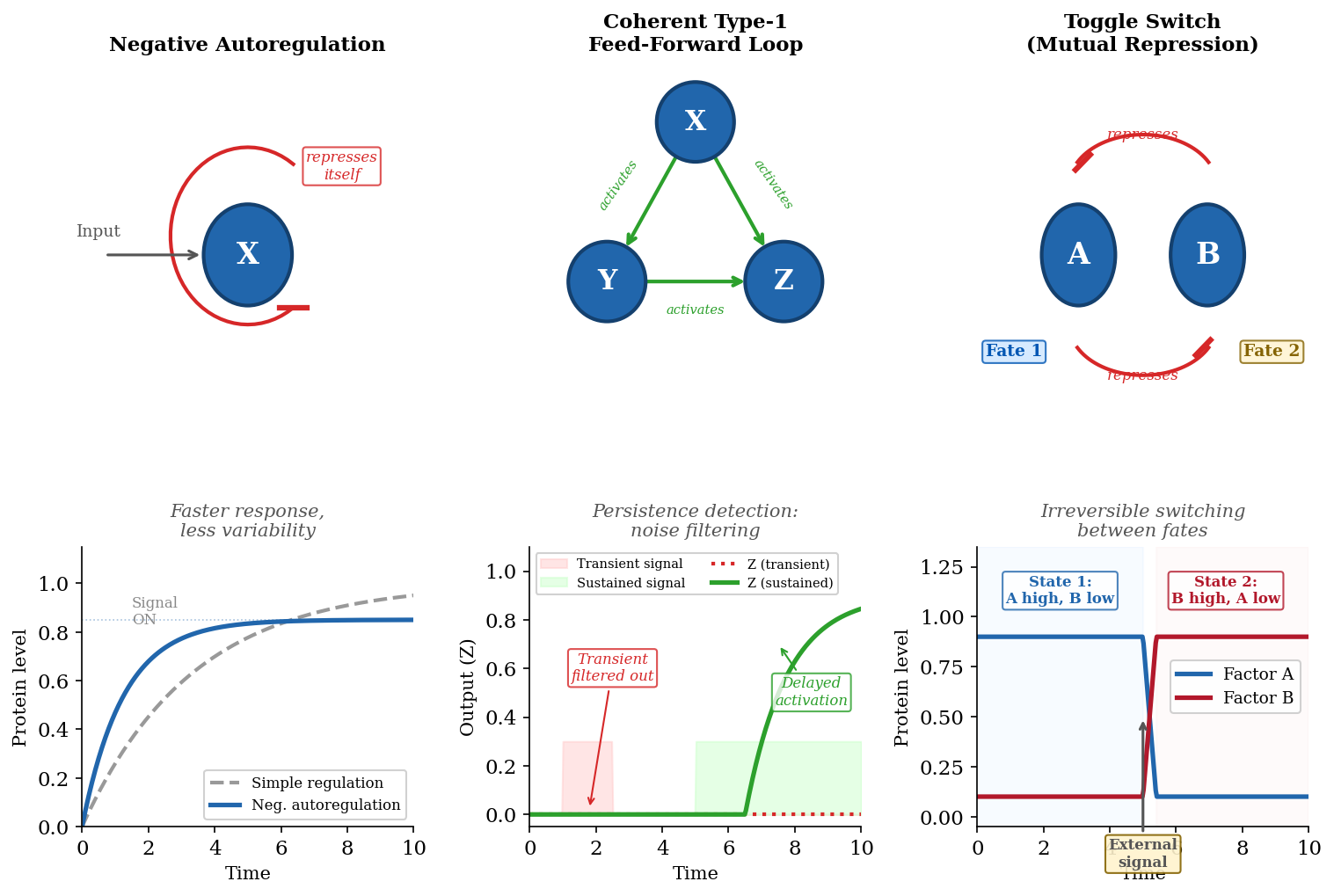
The systematic analysis of gene regulatory networks has revealed that certain small subgraph patterns, called network motifs, recur far more frequently than would be expected by chance. Uri Alon and colleagues, working initially in Escherichia coli, identified several motifs that appear to function as elementary computational units within regulatory networks. The simplest is autoregulation, in which a transcription factor regulates its own gene. Negative autoregulation — where a transcription factor represses its own transcription — is extremely common and speeds the response time of the circuit while reducing cell-to-cell variability in protein levels. Positive autoregulation — where a transcription factor activates its own transcription — is rarer but can generate bistability, locking the circuit into either a high-expression or low-expression state with a sharp transition between them.
Feed-Forward Loops and the Filtering of Noise
The feed-forward loop (FFL) is a three-gene motif in which transcription factor X regulates transcription factor Y, and both X and Y jointly regulate a target gene Z. The motif comes in eight varieties depending on whether each regulatory interaction is activating or repressing. The most common variant is the coherent type 1 FFL, in which X activates Y and both X and Y activate Z. This arrangement functions as a persistence detector: Z is activated only after X has been sustained long enough for Y to accumulate above its activation threshold. Transient pulses of X are filtered out because Y does not have time to reach the level needed to co-activate Z. The coherent type 1 FFL thus protects the cell against responding to noise in the input signal — a critical function in environments where stochastic fluctuations in transcription factor levels are unavoidable.
The incoherent type 1 FFL, in which X activates both Y and Z but Y represses Z, generates a pulse of Z expression followed by a return to baseline. This motif is found in pathways where a transient burst of gene expression is needed — for example, in the immediate-early response to growth factor stimulation, where genes such as c-FOS are rapidly and transiently induced. The pulse-generating property of the incoherent FFL arises because the direct activation of Z by X is initially unopposed, but as Y accumulates, its repressive effect on Z catches up and shuts the response down. The height and duration of the pulse can be tuned by adjusting the relative strengths and speeds of the direct and indirect arms of the loop.
Bistable Switches and Cell-Fate Decisions
Many biological decisions are irreversible: a cell that commits to becoming a neuron does not spontaneously revert to a stem cell under normal conditions. The network motif that underlies such irreversible decisions is the toggle switch, a two-gene circuit in which two transcription factors mutually repress each other. If factor A represses the gene encoding factor B, and factor B represses the gene encoding factor A, the system has two stable states: one in which A is high and B is low, and one in which B is high and A is low. An external signal that tips the balance toward one state will be self-reinforcing: if A gains the upper hand, it represses B, relieving repression of itself, which further increases A and further represses B, driving the system to the A-high/B-low steady state. Once there, the system resists perturbation — it is "locked in."
The paradigmatic example of a toggle switch in mammalian development is the mutual repression between the transcription factors GATA1 and PU.1 during haematopoietic differentiation. In a bipotent progenitor cell, both factors are expressed at moderate levels. Stochastic fluctuations or external signals that increase GATA1 repress PU.1, committing the cell to the erythroid (red blood cell) lineage. Conversely, signals that increase PU.1 repress GATA1, committing the cell to the myeloid (white blood cell) lineage. The mutual repression ensures that the decision is binary — intermediate states are unstable — and irreversible once the positive feedback has driven the system deep into one basin of attraction.
Oscillatory Circuits and Temporal Patterning
Not all regulatory networks settle into stable steady states. Some are designed to oscillate, producing rhythmic waves of gene expression that drive temporal patterning in development and physiology. The vertebrate segmentation clock, which generates the periodic somites (the precursors of vertebrae and ribs) during embryonic development, is driven by an oscillatory gene regulatory circuit involving the Notch, Wnt, and FGF signalling pathways. In the presomitic mesoderm, genes in the Hes/Her family undergo periodic cycles of activation and repression with a period of approximately two hours in mice. The oscillation arises from a delayed negative feedback loop: Hes7 protein represses its own transcription, but because protein synthesis, folding, and nuclear import take time, there is a delay between the onset of transcription and the accumulation of repressor. This delay creates a cycle in which Hes7 mRNA rises, Hes7 protein accumulates, repression kicks in, mRNA levels fall, protein decays, repression is relieved, and transcription resumes.
The circadian clock is an even more elaborate oscillatory circuit that generates approximately 24-hour rhythms in gene expression across virtually every tissue in the body. The core mechanism involves interlocking transcriptional-translational feedback loops: the transcription factors CLOCK and BMAL1 activate the genes encoding PER and CRY proteins, which accumulate in the cytoplasm, form complexes, translocate to the nucleus, and repress CLOCK-BMAL1-mediated transcription. As PER-CRY complexes are degraded (through phosphorylation by casein kinases and subsequent ubiquitin-mediated proteolysis), the repression is lifted and the cycle begins again. Superimposed on this core loop are accessory loops involving ROR and REV-ERB nuclear hormone receptors that stabilise the oscillation and modulate its amplitude. The precision of the circadian clock — it maintains a near-24-hour period even in the absence of external time cues — is a testament to the engineering sophistication achievable by interconnected gene regulatory circuits.
6. Morphogen Gradients and the Specification of Positional Identity
The regulatory mechanisms explored in the preceding sections — transcription factor control, signal transduction, post-transcriptional regulation, miRNA networks, and gene regulatory circuit logic — converge in one of the most elegant phenomena in biology: the specification of positional identity in a developing embryo by morphogen gradients. A morphogen is a signalling molecule that is produced at a localised source, diffuses through the tissue to form a concentration gradient, and activates different sets of target genes at different concentration thresholds. Cells at different positions along the gradient experience different morphogen concentrations, activate different transcription factor combinations, and consequently adopt different cell fates. The morphogen gradient thus converts a spatial cue (distance from the source) into a transcriptional programme (cell identity), using the regulatory logic of threshold responses, combinatorial control, and network motif topology.
The classic example is the Bicoid gradient in the Drosophila embryo. The mRNA encoding the transcription factor Bicoid is localised at the anterior (head) end of the egg, and after fertilisation, Bicoid protein diffuses toward the posterior, forming an exponentially decaying concentration gradient along the anterior-posterior axis. At high concentrations (near the head), Bicoid activates genes such as hunchback that specify anterior structures. At lower concentrations (farther from the head), Bicoid is insufficient to activate these targets, and posterior-specifying genes dominate instead. The sharpness of the hunchback expression boundary — far sharper than the smooth Bicoid gradient itself — is achieved by cooperative binding of Bicoid to multiple sites in the hunchback enhancer and by positive feedback in which Hunchback protein reinforces its own expression. The result is a near-digital switch from hunchback-on to hunchback-off at a precise position along the body axis, despite the inherently analogue nature of the diffusing morphogen.
Gradient Interpretation and the French Flag Model
Lewis Wolpert's French Flag model, proposed in 1969, provides a conceptual framework for understanding how morphogen gradients specify multiple cell fates. Imagine a row of cells exposed to a morphogen gradient. At high concentration, cells adopt fate A (blue); at intermediate concentration, fate B (white); at low concentration, fate C (red) — producing a tricolour pattern from a single gradient. The model predicts that the boundaries between fates are determined by the concentration thresholds at which specific transcription factors are activated, and that these thresholds are set by the binding affinities of the transcription factors for their target enhancers. A transcription factor with a high-affinity binding site will be activated at low morphogen concentrations (it responds to the long-range, low-concentration part of the gradient), while a factor with a low-affinity site will require high concentrations (it responds only near the source).
In vertebrate development, the Sonic Hedgehog (Shh) gradient in the developing neural tube provides a well-studied example. Shh is secreted from the notochord and floor plate at the ventral midline of the neural tube and forms a ventral-to-dorsal gradient. Different concentrations of Shh activate different combinations of homeodomain transcription factors — NKX2.2, OLIG2, NKX6.1, PAX6, IRX3, and DBX2 — each of which has a characteristic concentration threshold. The combinatorial expression of these factors divides the neural tube into discrete progenitor domains, each of which gives rise to a specific class of neurons (motor neurons, V0-V3 interneurons). Cross-repressive interactions between the transcription factors sharpen the domain boundaries, converting a smooth gradient into a series of discrete stripes — another instance of the toggle-switch motif operating at the tissue level.
Scaling, Robustness, and Self-Organisation in Gradient Systems
A striking feature of morphogen-mediated patterning is its robustness: the pattern of cell fates is remarkably insensitive to variations in embryo size, morphogen production rate, and temperature. In Drosophila, embryos that differ twofold in length still produce correctly proportioned body plans, indicating that the gradient and its readout scale with tissue size rather than being fixed at absolute concentration values. Several mechanisms contribute to this scaling. Expander molecules — secreted factors that are repressed by the morphogen itself — spread through the tissue and modulate morphogen diffusion or degradation, effectively stretching the gradient in proportion to the tissue domain. Feedback between the morphogen and its receptor can buffer the system against fluctuations in production rate: if morphogen levels rise, increased receptor-mediated endocytosis accelerates degradation, restoring the gradient to its normal shape. At the intracellular level, adaptation circuits in the signal transduction machinery allow cells to respond to the slope or fold-change in morphogen concentration rather than to its absolute level — a strategy that is inherently size-invariant. These self-organising properties mean that morphogen gradients are not fragile, top-down blueprints but resilient, feedback-stabilised systems that can pattern tissues reliably despite the noise and variability inherent in biological development.
From Gradients to Gene Regulatory Networks: Integrating the Layers
The specification of cell fate by morphogen gradients is not a simple readout of concentration. In reality, cells integrate the morphogen signal with their temporal history (how long they have been exposed), with signals from other pathways (Wnt, BMP, FGF), with their epigenetic state (which enhancers are accessible in chromatin), and with the miRNA landscape that tunes the levels of the responding transcription factors. The Shh gradient in the neural tube, for example, is interpreted differently at different developmental stages — early exposure to a given concentration produces a different fate than late exposure to the same concentration — because the chromatin landscape and the repertoire of expressed transcription factors change over time.
This temporal dimension connects the instantaneous logic of signalling pathways to longer-term cellular memory. The initial morphogen signal may activate a transcription factor that recruits histone acetyltransferases to a set of enhancers, making those enhancers accessible for subsequent transcription factor binding even after the morphogen signal has faded. In this way, a transient signalling event is converted into a stable epigenetic change — a cell-fate decision that persists through subsequent cell divisions. The gene regulatory networks described here are not isolated circuits operating in a vacuum; they are embedded in a chromatin landscape that remembers their past activity and constrains their future responses. That landscape — the histone code, the enhancer repertoire, and the three-dimensional genome organisation — and its interplay with the network logic explored here are among the central themes of modern gene regulation.